File:Tubifera dudkae-4.jpg
Jump to navigation
Jump to search


Size of this preview: 406 × 599 pixels. Other resolutions: 162 × 240 pixels | 325 × 480 pixels | 406 × 600 pixels | 520 × 768 pixels | 694 × 1,024 pixels | 2,134 × 3,150 pixels.
Original file (2,134 × 3,150 pixels, file size: 3.15 MB, MIME type: image/jpeg)
Summary[edit]
| Description |
Українська: Внутрішня оболонка спорангію рідкісного міксоміцета Tubifera dudkae вкрита складками, що нагадують морські хвилі. Серед них трапляються сітчасті спори. Мікрофотографія виконана за допомогою растрового електронного мікроскопу, кольори одержані засобами комп’ютерної графіки. Збільшення х2000
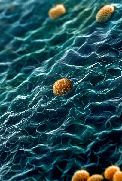
English: Internal surface of the peridium of the rare myxomycete Tubifera dudkae is covered with folds, resembling sea waves. Among them oval shaped reticulate spores occur. Micrograph image made with a scanning electronic microscope and coloured with computer graphic tools. Magnification 2000x.
Русский: Внутренняя оболочка пиридия редкого миксомицета Tubifera dudkae покрыта складками, напоминающими морские волны. Среди них встречаются сетчатые споры. Микрофотография выполнена с помощью сканирующего электронного микроскопа, цвета добавлены средствами компьютерной графики. Увеличение х2000
Español: La superficie interna del peridio de Tubifera dudkae, un hongo mucilaginoso de la clase de los mixomicetos, está cubierta con pliegues algo semejantes a las olas del mar. Entre ellos se encuentran esporas reticulares de forma oval. Imagen de micrografía, con una ampliación de 2000x, obtenida con un microscopio electrónico de barrido y coloreada con software gráfico.
Português: Fotomicrografia com uma ampliação de 2000x mostra a superfície interna do perídio do raro Tubifera dudkae, um fungo mucilaginoso da classe Myxogastria. Está coberta por dobras, que se assemelham a ondas do mar e entre elas se encontram esporos reticulares de forma oval. Български: Вътрешната повърхност на перидиума (защитната обвивка) на рядката миксомицета (лигава гъба) Тубифера (Tubifera dudkae) е покрита с гънки, наподобяващи морски вълни. Между тях се забелязват овалните тела на мрежестите спори. Микроскопско изображение с увеличение 2000x
Polski: Mikrofotografia wykonana przy powiększeniu 2000× przedstawiająca wewnętrzną powierzchnię perydium rzadkiego śluzowca z gatunku Tubifera dudkae pokrytą fałdami przypominającymi trochę morskie fale, wśród których występują owalne zarodniki o siatkowatej powierzchni.
|
| Date | |
| Source | Own work |
| Author | Дмитро Леонтьєв |
| This image was uploaded as part of European Science Photo Competition 2015. |
Assessment[edit]
|
| This image was selected as picture of the day on Wikimedia Commons for . It was captioned as follows: English: The internal surface of the peridium of the rare myxomycete Tubifera dudkae, a plasmodial slime mold, is covered with folds somewhat resembling sea waves. Among them oval shaped reticulate spores occur. Micrograph image with a magnification of 2000x. Čeština: Vnitřní povrch tkáně peridia vzácných organizmů vlastní hlenky (tubifera) poněkud připomíná mořské vlny, mezi nimiž jsou vidět spóry oválného tvaru. Mikrofotografie se zvětšením 2000x. English: The internal surface of the peridium of the rare myxomycete Tubifera dudkae, a plasmodial slime mold, is covered with folds somewhat resembling sea waves. Among them oval shaped reticulate spores occur. Micrograph image with a magnification of 2000x. Español: La superficie interna del peridio de Tubifera dudkae, un hongo mucilaginoso de la clase de los mixomicetos, está cubierta con pliegues algo semejantes a las olas del mar. Entre ellos se encuentran esporas reticulares de forma oval. Imagen de micrografía con una ampliación de 2000x. Français: La surface interne du péridium du rare myxomycète Tubifera dudkae, couverte de plis semblables à des vagues, avec quelques spores ovales jaunes. Image réalisée par photomicrographie avec un grandissement de 2000:1. Magyar: Egy ritka nyálkagomba, a Tubifera dudkae perídiumának felülete a spórákkal (elektronmikroszkópos felvétel) Nederlands: De binnenzijde van het peridium van de zeldzame Tubifera dudkae, een echte slijmzwam, doet enigszins aan golven denken. Op de golven 'drijven' ovale sporen. Vergroting: 2000× Polski: Mikrofotografia wykonana przy powiększeniu 2000× przedstawiająca wewnętrzną powierzchnię perydium rzadkiego śluzowca z gatunku Tubifera dudkae pokrytą fałdami przypominającymi trochę morskie fale, wśród których występują owalne zarodniki o siatkowatej powierzchni. Português: Fotomicrografia com uma ampliação de 2000x mostra a superfície interna do perídio do raro Tubifera dudkae, um fungo mucilaginoso da classe Myxogastria. Está coberta por dobras, que se assemelham a ondas do mar e entre elas se encontram esporos reticulares de forma oval. Български: Вътрешната повърхност на перидиума (защитната обвивка) на рядката миксомицета (лигава гъба) Тубифера (Tubifera dudkae) е покрита с гънки, наподобяващи морски вълни. Между тях се забелязват овалните тела на мрежестите спори. Микроскопско изображение с увеличение 2000x |
Licensing[edit]
I, the copyright holder of this work, hereby publish it under the following license:
|
This file is licensed under the Creative Commons Attribution-Share Alike 4.0 International license. | |
|
File history
Click on a date/time to view the file as it appeared at that time.
| Date/Time | Thumbnail | Dimensions | User | Comment | |
|---|---|---|---|---|---|
| current | 10:24, 29 October 2015 | | 2,134 × 3,150 (3.15 MB) | Дмитро Леонтьєв (talk | contribs) | User created page with UploadWizard |
- You cannot overwrite this file.
File usage on Commons
The following 94 pages uses this file:
- User:Daniel Mietchen/POTY/2016
- User:OgreBot/Uploads by new users/2015 October 29 09:00
- User:Sophie Österberg (WMSE)/nyflip
- User talk:Kruusamägi
- User talk:Дмитро Леонтьєв
- Commons:European Science Photo Competition 2015/Winners
- Commons:European Science Photo Competition 2015/finalists/Microscopy images
- Commons:European Science Photo Competition 2015 in Ukraine
- Commons:Featured picture candidates/File:Tubifera dudkae-4.jpg
- Commons:Featured picture candidates/Log/May 2016
- Commons:Featured pictures
- Commons:Featured pictures, list
- Commons:Featured pictures/Other lifeforms
- Commons:Featured pictures/af
- Commons:Featured pictures/als
- Commons:Featured pictures/an
- Commons:Featured pictures/ar
- Commons:Featured pictures/ast
- Commons:Featured pictures/be-tarask
- Commons:Featured pictures/bn
- Commons:Featured pictures/br
- Commons:Featured pictures/ca
- Commons:Featured pictures/chronological/2016-A
- Commons:Featured pictures/cs
- Commons:Featured pictures/da
- Commons:Featured pictures/de
- Commons:Featured pictures/el
- Commons:Featured pictures/eo
- Commons:Featured pictures/es
- Commons:Featured pictures/eu
- Commons:Featured pictures/ext
- Commons:Featured pictures/fi
- Commons:Featured pictures/fr
- Commons:Featured pictures/gl
- Commons:Featured pictures/gu
- Commons:Featured pictures/he
- Commons:Featured pictures/hr
- Commons:Featured pictures/hy
- Commons:Featured pictures/ia
- Commons:Featured pictures/id
- Commons:Featured pictures/it
- Commons:Featured pictures/ko
- Commons:Featured pictures/lb
- Commons:Featured pictures/mk
- Commons:Featured pictures/ml
- Commons:Featured pictures/mo
- Commons:Featured pictures/ms
- Commons:Featured pictures/nan
- Commons:Featured pictures/nl
- Commons:Featured pictures/no
- Commons:Featured pictures/oc
- Commons:Featured pictures/pl
- Commons:Featured pictures/pt
- Commons:Featured pictures/ro
- Commons:Featured pictures/ru
- Commons:Featured pictures/sk
- Commons:Featured pictures/sl
- Commons:Featured pictures/sq
- Commons:Featured pictures/su
- Commons:Featured pictures/sv
- Commons:Featured pictures/uk
- Commons:Featured pictures/vi
- Commons:Imatges de qualitat
- Commons:Mmàggini n vitrina
- Commons:Picture of the Year/2016/Candidates
- Commons:Picture of the Year/2016/R1/Gallery/2016-A
- Commons:Picture of the Year/2016/R1/Gallery/ALL
- Commons:Picture of the Year/2016/R1/Gallery/M05
- Commons:Picture of the Year/2016/R1/Gallery/Objects, shells and miscellaneous
- Commons:Picture of the Year/2016/R1/v/Tubifera dudkae-4.jpg
- Commons:Science Photo Competition 2016 in Ukraine
- Commons:Science Photo Competition 2016 in Ukraine/Image categories
- Commons:Seçilmiş şəkil
- Commons:Seçkin resimler/tr
- Commons:Wiki Science Competition 2017 in Ukraine
- Commons:Wiki Science Competition 2017 in Ukraine/Категорії зображень
- Commons:រូបភាពពិសេស/km
- Commons:正圖
- Commons:特色图片
- Commons:特色圖片
- Commons:特色靚相
- Commons:秀逸な画像
- Template:Potd/2017-06
- Template:Potd/2017-06-09
- Template:Potd/2017-06-09 (bg)
- Template:Potd/2017-06-09 (cs)
- Template:Potd/2017-06-09 (en)
- Template:Potd/2017-06-09 (es)
- Template:Potd/2017-06-09 (fr)
- Template:Potd/2017-06-09 (hu)
- Template:Potd/2017-06-09 (mk)
- Template:Potd/2017-06-09 (nl)
- Template:Potd/2017-06-09 (pl)
- Template:Potd/2017-06-09 (pt)
File usage on other wikis
The following other wikis use this file:
- Usage on be-tarask.wikipedia.org
- Usage on ca.wikipedia.org
- Usage on crh.wikipedia.org
- Usage on cv.wikipedia.org
- Usage on en.wikipedia.org
- Usage on es.wikipedia.org
- Usage on hu.wikipedia.org
- Usage on hy.wikipedia.org
- Usage on it.wikipedia.org
- Usage on ka.wikipedia.org
- Usage on ko.wikipedia.org
- Usage on krc.wikipedia.org
- Usage on lbe.wikipedia.org
- Usage on lez.wikipedia.org
- Usage on mdf.wikipedia.org
- Usage on mk.wikipedia.org
- Usage on os.wikipedia.org
- Usage on pt.wikipedia.org
- Usage on ru.wikipedia.org
- Usage on sah.wikipedia.org
- Usage on sq.wikipedia.org
- Usage on tyv.wikipedia.org
- Usage on udm.wikipedia.org
- Usage on uk.wikipedia.org
- Usage on vi.wikipedia.org
- Usage on xal.wikipedia.org
- Usage on zh.wikipedia.org